▎Glutathione Research
Molecular Background
Glutathione (GSH), also known as L-γ-glutamyl-L-cysteinylglycine, is a tripeptide composed of glutamate, cysteine, and glycine. Unlike a conventional α-peptide linkage, the glutamate residue is connected to cysteine through its γ-carboxyl group. The molecule contains a cysteinyl thiol (-SH), which is the principal redox-active group in its structure. PubChem lists reduced glutathione under CID 124886, with the molecular formula C10H17N3O6S and a molecular weight of 307.33 g/mol.[6]
Research Progress
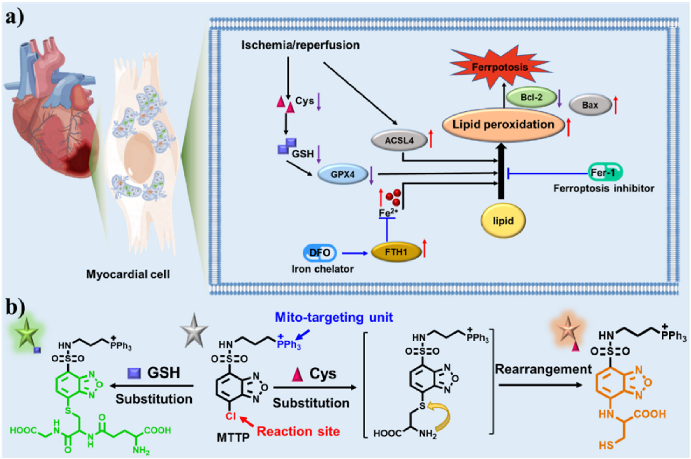
Zeng Jiayu and colleagues developed a mitochondria-targeted fluorescent probe (MTTP) capable of distinguishing cysteine and glutathione (GSH) through two different emission signals. The probe was used to monitor changes in mitochondrial cysteine and GSH during ferroptosis and its inhibition. In an oxygen-glucose deprivation/reoxygenation (OGD/R) model using H9C2 cells, the authors applied MTTP to visualize changes in these thiols under experimental conditions associated with myocardial ischemia/reperfusion injury.[1]
![Fig1]()
Fig1. The graphic mechanism for monitoring ferroptosis in OGD/R-induced MI/R injury via MTTP[1].
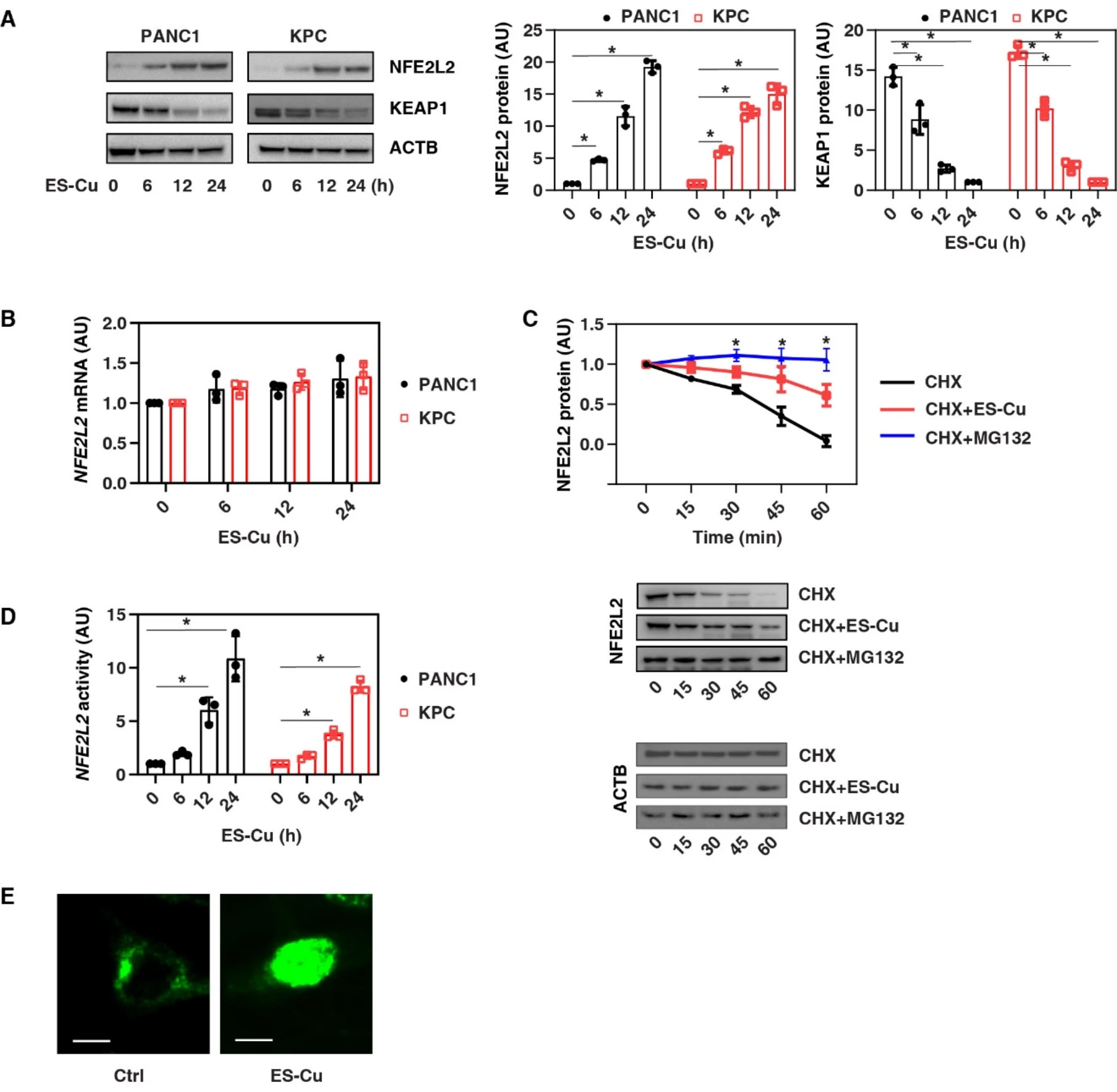
Jiao Liu et al. investigated the role of GSH in cuproptosis using pancreatic ductal adenocarcinoma (PDAC) cell lines and mouse tumor models. The authors reported that GSH chelated copper through its thiol group and reduced mitochondrial copper-associated proteotoxic stress under the experimental conditions. Their mechanistic experiments showed that ES-Cu increased NFE2L2 protein stability and upregulated GSH-synthesis-related genes, while SLC25A39 was involved in transporting GSH into mitochondria. Genetic inhibition of the NFE2L2-GSH-SLC25A39 pathway increased cuproptosis-associated responses in the cell and mouse models studied.[2]
![Fig2]()
Fig2. Enhanced NFE2L2 protein stability and activity during cuproptosis[2].
(A, B) Western blot and qPCR analyses of the expression of NFE2L2 in PDAC cells in whole cell extracts after treatment with ES-Cu (100 nM, 1:1) for 6–24 h (n = 3 biologically independent samples, *P < 0.05 versus control, ANOVA test). (C) CHX chase assay of the degradation kinetics of NFE2L2 protein in PANC1 cells after treatment with CHX (50 µg/ml) in the absence or presence of 100 nM ES-Cu or 2 µM MG132 for 15–60 min (n = 3 biologically independent samples, *P < 0.05 versus CHX group, ANOVA test). ACTB was used as a loading control. (D) NFE2L2 transcriptional activity assay of PDAC cells following treatment with ES-Cu (100 nM) for 6–24 h (n = 3 biologically independent samples, *P < 0.05 versus control, ANOVA test). (E) Image analysis of the localization of NFE2L2 in PANC1 cells after treatment with ES-Cu for 24 h. Bar = 15 μm.
Gurung et al. investigated glutathione metabolism in argininosuccinic aciduria (ASA) using targeted metabolomics in individuals with ASL deficiency and in mouse models. The authors reported glutathione depletion together with altered upstream cysteine utilization and changes in antioxidant pathways. In the preclinical mouse studies, lipid-nanoparticle-encapsulated human ASL mRNA improved ASL activity, ureagenesis, and measured glutathione-metabolism endpoints. The intervention evaluated in this study was ASL mRNA therapy rather than administration of exogenous glutathione.[3]
Wang et al. prepared palladium nanocrystals by microwave synthesis and developed a nanoenzyme-based colorimetric method for GSH determination. The assay used the interaction between the GSH thiol group and palladium nanocrystals to alter the catalytic color reaction. The authors reported linear detection ranges of 3-40 μM and 55-110 μM, with a detection limit of 0.14 μM. The method was also evaluated in serum samples, with recovery values reported in the original study.[4]
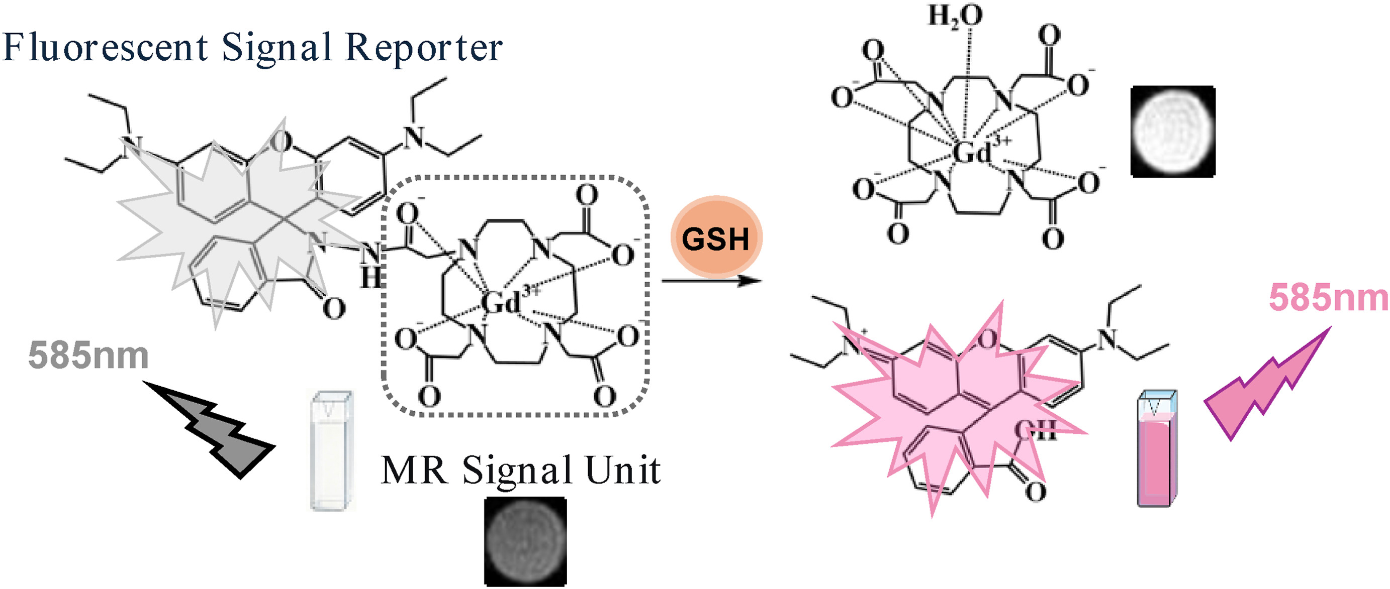
Chen et al. developed a rhodamine-Gd(III)-based fluorescent/magnetic resonance dual-modal probe (RG) for GSH detection. In the presence of GSH, the probe generated coordinated fluorescence and magnetic resonance signal changes. The authors reported a response time of approximately 5 ms and a detection limit of 2.47 μM, and subsequently applied the probe to dual-modal GSH imaging in a mouse model.[5]
![Fig3]()
Fig3. Design strategy of probe RG for GSH detection.
Research Limitations
The studies summarized above address different research questions, including fluorescent sensing, cell-death mechanisms, inherited metabolic disease, colorimetric quantification, and imaging-probe development. Most of these studies measured endogenous GSH or used GSH as an analytical target rather than evaluating an externally supplied glutathione material. Results obtained in H9C2 cells, PDAC models, ASL-deficient subjects or mice, analytical serum samples, and imaging models are therefore not directly interchangeable.[1-5]
Reduced glutathione is the chemical entity identified in PubChem as CID 124886.[6] Experimental findings in the cited literature do not establish the identity, purity, safety, or clinical performance of a separately supplied research material. Interpretation should remain tied to the exact model, analytical method, and material described in each source.
COCER Peptides presents Glutathione solely for laboratory research and analytical use. It is not intended for human or veterinary use. The research described above summarizes findings reported in third-party publications and is not a statement of clinical efficacy, safety, diagnosis, treatment, prevention, or an instruction for use.