▎GLP-2 Research
Research Progress
Anthony P. Ramsanahie and colleagues used the sodium/glucose cotransporter 1 (SGLT1) as a marker to investigate the effects of long-term [Gly2]GLP-2 administration on diurnal gene-expression rhythms in rats. Animals received [Gly2]GLP-2 twice daily for 10 days, while the control group received vehicle. SGLT1 mRNA expression was assessed by Northern blot hybridization and in situ hybridization, and SGLT1 protein was evaluated by immunofluorescence staining and Western blotting. The authors reported increased villus height, crypt depth, and proliferation index in the [Gly2]GLP-2 group. The diurnal upward trend in SGLT1, GLUT2, and GLUT5 mRNA was not altered, while SGLT1 protein abundance was higher at the ZT3 and ZT9 time points and showed greater localization at the apical membrane under the study conditions.[1]
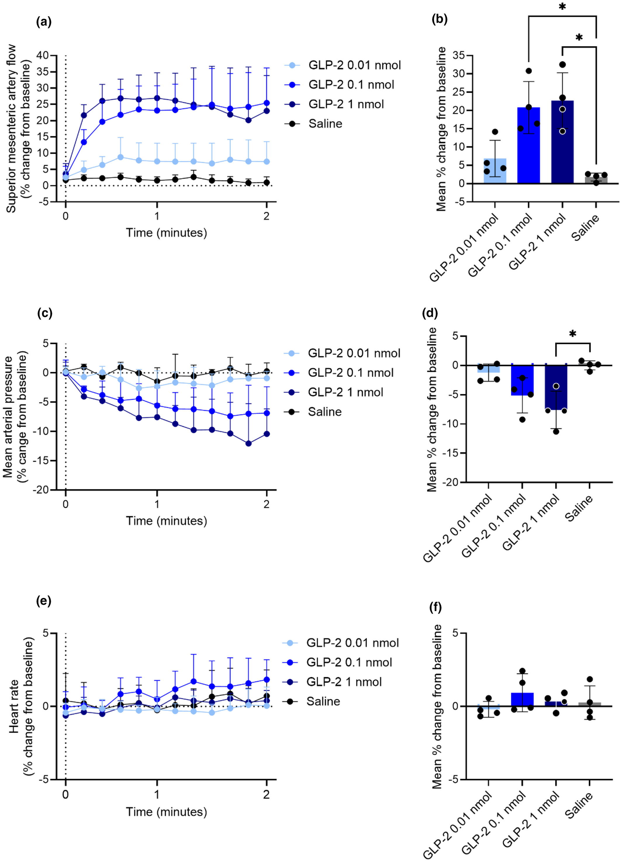
Katrine D. Galsgaard and colleagues used a transit-time blood-flow probe to examine the acute effects of GLP-2 and GIP, alone and in combination, on superior mesenteric artery blood flow in anesthetized male rats. The investigators also tested nitric oxide synthase inhibition, vasoactive intestinal peptide receptor antagonism, a GIP/GLP-2 receptor co-agonist, and the GLP-2 receptor antagonist GLP-2(3-33). The authors reported acute increases in superior mesenteric artery blood flow after GLP-2, GIP, and co-agonist exposure. In this model, the GLP-2-associated blood-flow response was not prevented by nitric oxide synthase inhibition or VIP receptor antagonism, while GLP-2(3-33) antagonized the response to GLP-2.[2]
![Fig1]()
Fig1. Acute changes in superior mesenteric artery blood flow reported after GLP-2 exposure in anesthetized rats.[2]
Percent change from baseline in (a and b) superior mesenteric artery blood flow, (c and d) mean arterial blood pressure, and (e and f) heart rate after intravenous bolus administration of study-specified doses of GLP-2 or saline. Statistical methods, group size, and dose information are reported in the original publication.[2]
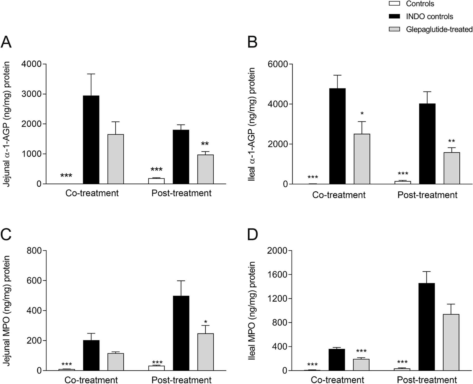
Jolanta Skarbaliene and colleagues induced small-intestinal inflammation in Wistar rats with indomethacin and studied glepaglutide, a long-acting GLP-2 receptor agonist, using co-treatment and post-treatment regimens. Small-intestinal length and the inflammatory markers α-1-acid glycoprotein and myeloperoxidase were measured, while small-intestinal mass was used as an enterotrophic endpoint. The authors reported differences in intestinal length, inflammatory-marker concentrations, and intestinal mass in the glepaglutide study groups compared with indomethacin controls. These findings relate specifically to glepaglutide and the experimental rat model described in the publication, rather than to native GLP-2 as an interchangeable research material.[3]
![Fig2]()
Fig2. Jejunal and ileal inflammatory-marker measurements reported after glepaglutide co-treatment or post-treatment in an indomethacin-induced rat model of small-intestinal inflammation.[3]
The source study evaluated glepaglutide at the study-specified dose in co-treatment and post-treatment regimens. Jejunal and ileal α-1-acid glycoprotein and myeloperoxidase concentrations were measured during the active inflammation phase. Group definitions, statistical comparisons, and complete dosing details are reported in the original publication.[3]
Jie Wu and colleagues encapsulated porcine GLP-2 (pGLP-2) in microspheres and examined the formulation in dextran sulfate sodium (DSS)-treated mice. The authors reported an initial burst release of 20.36% followed by sustained release for at least 9 days. In the DSS mouse model, the pGLP-2 microsphere group showed differences in body weight, colon length, small-intestinal weight, colon-injury scores, Occludin mRNA, and the measured inflammatory cytokine transcripts compared with the DSS control groups.[4]
Xia Chen and colleagues randomly assigned 30 rats to a sham group or to groups in which severe acute pancreatitis was induced by retrograde pancreatico-biliary duct injection of sodium taurocholate. One of the pancreatitis groups received GLP-2 under the study protocol. Intestinal-barrier changes were evaluated by histology, plasma diamine oxidase concentrations, proliferating cell nuclear antigen staining, and epithelial-cell apoptosis. The authors reported differences in ileal histological scores, diamine oxidase concentrations, apoptosis, and proliferating cell nuclear antigen staining in the GLP-2 study group compared with the untreated pancreatitis group.[5]
Paul A. Kitchen and colleagues established a parenteral-nutrition (PN) rat model and administered GLP-2, epidermal growth factor (EGF), or the combination for 6 days. Compared with the untreated PN group, the authors reported increases in small-intestinal weight of 75%, 43%, and 116% in the GLP-2, EGF, and GLP-2+EGF groups, respectively, and increases in proximal small-intestinal cell proliferation of 2.3-fold, 1.7-fold, and 3.4-fold. The combined GLP-2+EGF group also showed a greater change in villus and crypt area than either factor alone under the study conditions. GLP-2 did not produce the same colon response reported for EGF, and the combination group showed changes in distal small-intestinal IBABP transcript expression.[6]
Kundanika Mukherjee and colleagues examined whether gut-brain neural communication contributed to GLP-2-associated post-absorptive intestinal lipid mobilization in rats with mesenteric lymphatic cannulation. A complete subdiaphragmatic vagotomy was used to disrupt gut-brain communication, and lymphatic lipid output was measured after a lipid load following intraperitoneal GLP-2 or saline exposure. The authors reported increased lymphatic lipid output and activation of proopiomelanocortin neurons after peripheral GLP-2 exposure, while vagotomy reduced the GLP-2-associated change in intestinal lipid release.[7]
Research Limitations
The studies summarized above were conducted in rats or mice and used different experimental systems, exposure conditions, endpoints, and research materials. [Gly2]GLP-2, native GLP-2, porcine GLP-2, and glepaglutide are not the same molecular entity and should not be treated as interchangeable when comparing study results. Findings from anesthetized-animal, inflammatory-disease, parenteral-nutrition, and surgically modified models remain specific to the conditions in which they were generated.[1-7]
The cited literature does not establish that observations made in these animal models predict safety, efficacy, or physiological effects in humans. Differences in peptide sequence, formulation, route of exposure, study duration, species, and tissue context can materially affect experimental results. For research interpretation, conclusions should therefore remain tied to the exact peptide or analogue and model reported in each source.
COCER Peptides presents this material for laboratory research use only. It is not intended for human or veterinary use. The research described above summarizes findings reported in third-party publications and is not a statement of clinical efficacy, safety, diagnosis, treatment, prevention, or an instruction for use.