
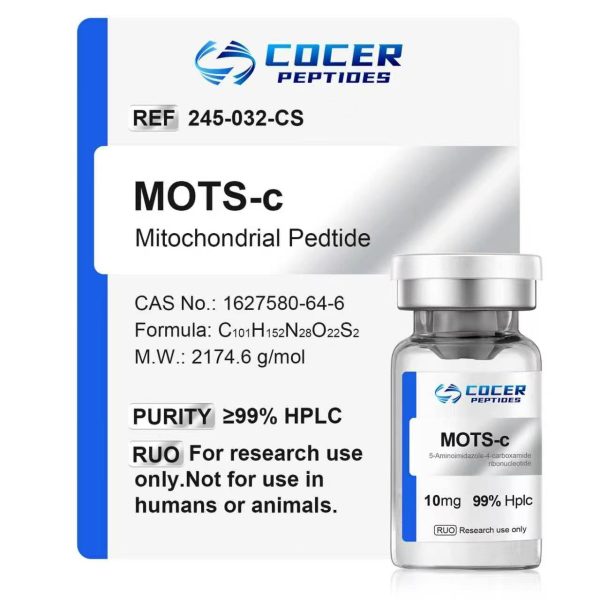
MOTs-C 10mg
MOTS-c is a mitochondrial-derived 16-amino-acid peptide encoded within the MT-RNR1 region of mitochondrial DNA. Supplied as a lyophilized powder for laboratory research and analytical use only. Not intended for human or veterinary use, including consumption, injection, topical application, or any diagnostic, therapeutic, or clinical purpose.
Certificate of Analysis (COA)
Click “Check the COA” in the product section above to access the available Certificate of Analysis for this product. Please refer to the report for batch information and analytical results.
For laboratory research use only. Not for human or veterinary use.
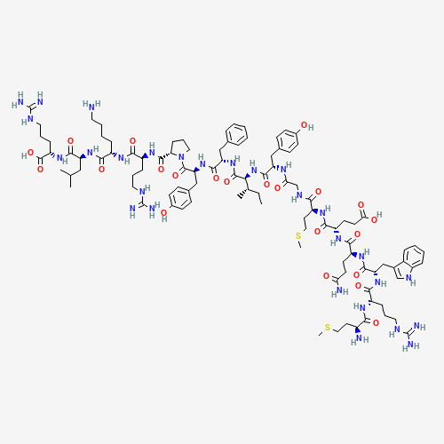
MOTs-CStructure
 PubChem
- Sequence:MRWQEMGYIFYPRKLR
- Molecular Formula:C101H152N28O22S2
- Molecular Weight:2174.6g/mol
- CAS Number:1627580-64-6
- PubChem CID:146675088
MOTs-C Research
Molecular Background
Mitochondrial open reading frame of the 12S rRNA-c (MOTS-c) is a 16-amino-acid mitochondrial-derived peptide encoded by a short open reading frame within the mitochondrial 12S rRNA region (MT-RNR1). PubChem lists the sequence as MRWQEMGYIFYPRKLR, with PubChem CID 146675088, molecular formula C101H152N28O22S2, and a molecular weight of 2174.6 g/mol.[1,8]
Research Progress
In an in vitro study, Jingsong Xiao and colleagues examined the expression and localization of MOTS-c in PC12 cells exposed to rotenone. The authors observed increased translocation of MOTS-c from mitochondria to the nucleus and reported an interaction with Nrf2 associated with changes in HO-1 and NQO1 expression. In accompanying cell and rat experiments, MOTS-c pretreatment was associated with differences in markers of mitochondrial dysfunction, oxidative stress, and the striatal expression of TH, PSD95, SYP, Nrf2, HO-1, NQO1, and Keap1 under rotenone exposure.[2]
Fig1. MOTS-c expression and subcellular localization measured in PC12 cells exposed to rotenone in the cited study.[2]
A, MOTS-c protein levels in whole-cell samples. B, MOTS-c levels in culture supernatant measured by ELISA. C, MOTS-c levels measured in mitochondrial, nuclear, and cytoplasmic fractions of PC12 cells. Values are presented as mean ± SEM in the source study; *P < 0.05 and **P < 0.01 versus the control group.[2]
Jingwei Shi and colleagues studied MOTS-c in experimental ischemic flap models and used RNA sequencing, tissue clearing, laser speckle contrast imaging, Doppler analysis, histological staining, Western blotting, ELISA, and immunofluorescence to examine perfusion, vascular and collagen-related changes, autophagy, pyroptosis, lysosomal membrane permeabilization (LMP), and associated signaling pathways. The authors reported higher blood-flow measurements and changes in angiogenesis and collagen remodeling in the MOTS-c groups. Their mechanistic experiments also linked the observed responses to PLA2G4A phosphorylation and the MAPK1-MAPK3-NF-κB signaling cascade under the conditions of the model.[3]
Fig2. Perfusion, tissue, and pathway-related findings reported in the ischemic flap model studied by Shi et al.[3]
(A-C) On postoperative day 7, the source study compared flap appearance, survival area, and subcutaneous blood-flow signals among sham, flap, and MOTS-c study groups. (D-H) Tissue-clearing images, oxygenation and blood-flow measurements, HE and Masson staining, vessel density, and collagen volume fraction were evaluated. (I-K) RNA-sequencing data were analyzed using KEGG and GO enrichment methods. Statistical methods and group sizes are reported in the original publication.[3]
Yanli Zhang and colleagues examined MOTS-c in a C57BL/6 mouse model of radiation-induced lung injury and in MLE-12 and primary mouse lung epithelial cell experiments. Following pulmonary irradiation, the authors reported differences in histological lung-injury scores, inflammatory and oxidative-stress markers, and mitochondrial-associated endpoints in the MOTS-c study groups. They also observed increased Nrf2 levels and nuclear translocation, and experiments involving Nrf2 deficiency supported an Nrf2-dependent component of the responses reported in this model.[4]
Fig3. Histological, inflammatory, oxidative-stress, and Nrf2-related measurements reported in the irradiated mouse model.[4]
(A-E) The source study shows the experimental timelines for pulmonary irradiation and MOTS-c study groups, together with MOTS-c measurements in lung tissue and serum. (F-Q) Lung histology, BALF measurements, serum oxidative-stress markers, and IL-6 and TNF-α mRNA were evaluated. Group sizes, study doses, statistical comparisons, and experimental procedures are provided in the original publication.[4]
Saranya Sri Santhanam and colleagues used isolated Langendorff-perfused rat hearts subjected to 30 minutes of global ischemia followed by 60 minutes of reperfusion. During early reperfusion, the isolated-heart preparations were exposed to study-specified MOTS-c doses through Krebs-Henseleit buffer. The investigators measured hemodynamic variables, infarct area, lactate dehydrogenase release, oxidative-stress markers, inflammatory measures, and apoptosis-related gene expression. Within the tested range, the authors reported the largest change at 0.5 mg/kg, including a 73% lower infarct area and 65% lower lactate dehydrogenase release compared with the ischemia-reperfusion group. The study also reported changes in antioxidant enzymes, lipid peroxidation, myeloperoxidase activity, apoptosis-related genes, and in silico peptide-protein interactions. Because the work used an isolated-heart preparation, the authors noted that systemic influences and pharmacokinetic factors were not represented.[5]
Fig4. Experimental workflow for the isolated rat-heart ischemia-reperfusion model, including heart isolation, Langendorff perfusion, stabilization, ischemia, reperfusion, and the study groups evaluated by Santhanam et al.[5]
Zhe Wang and colleagues used 8- to 10-week-old male Kunming mice to establish acute and chronic inflammatory pain models using formalin, capsaicin, λ-carrageenan, and complete Freund’s adjuvant (CFA). Behavioral testing, qPCR, Western blotting, ELISA, immunohistochemistry, and immunofluorescence were used to examine nociceptive responses and inflammatory signaling. The authors reported lower behavioral hypersensitivity responses in the MOTS-c study groups and changes in inflammatory mediators, glial and neuronal activation in the spinal dorsal horn, and peripheral CGRP- and P2X3-positive neuronal signaling. These findings were obtained in mouse models and were not clinical observations.[6]
Research Limitations
The studies summarized above use different experimental systems, including cultured cells, rodent models, isolated rat hearts, and disease-specific animal models. Results from these systems are not directly interchangeable, and differences in species, tissue environment, exposure conditions, endpoints, and study design can affect the observations reported. A 2024 systematic review and meta-analysis of circulating mitochondrial-derived peptides also reported substantial heterogeneity among the available studies, limited sample sizes, variation in assay methods and sample types, and an insufficient number of studies for several subgroup analyses.[7] These limitations restrict direct translation of the current literature to human use.
COCER Peptides provides MOTS-c solely as a laboratory research material. It is not intended for human or veterinary use, administration, diagnosis, treatment, prevention, or any other clinical application. The research described on this page is drawn from third-party publications and is presented only to document reported experimental findings; it does not represent a statement of clinical safety or efficacy for any COCER Peptides material.
Relevant Citations
[1] Zheng Y, Wei Z, Wang T. MOTS-c: A promising mitochondrial-derived peptide for therapeutic exploitation[J]. Frontiers in Endocrinology, 2023,14:1120533.
DOI:10.3389/fendo.2023.1120533.
[2] Xiao J, Zhang Q, Shan Y, et al. The Mitochondrial-Derived Peptide (MOTS-c) Interacted with Nrf2 to Defend the Antioxidant System to Protect Dopaminergic Neurons Against Rotenone Exposure[J]. Molecular Neurobiology, 2023,60(10):5915-5930.
DOI:10.1007/s12035-023-03443-3.
[3] Shi J, Wu Y, Liu X, et al. MOTS-c, a mitochondrial-derived peptide, ameliorates lysosomal membrane permeability and improves survival of soft tissue transplantation[J]. Autophagy, 2026,22(10):2695-2724.
DOI:10.1080/15548627.2026.2677180.
[4] Zhang Y, Huang J, Zhang Y, et al. The Mitochondrial-Derived Peptide MOTS-c Alleviates Radiation Pneumonitis via an Nrf2-Dependent Mechanism[J]. Antioxidants, 2024,13(5):613.
DOI:10.3390/antiox13050613.
[5] Santhanam S S, Jayaraman S, Kurian G A. Exogenous MOTS-c mitigates myocardial ischemia–reperfusion injury: experimental and in silico evidence from rat heart models[J]. Naunyn-Schmiedeberg’s Archives of Pharmacology, 2026,399(7):9969-9984.
DOI:10.1007/s00210-026-05018-0.
[6] Wang Z, Yang L, Xu L, et al. Central and peripheral mechanism of MOTS-c attenuates pain hypersensitivity in a mice model of inflammatory pain[J]. Neurological Research, 2024,46(2):165-177.
DOI:10.1080/01616412.2023.2258584.
[7] Zhou Q, Yin S, Lei X, et al. The correlation between mitochondrial derived peptide (MDP) and metabolic states: a systematic review and meta-analysis[J]. Diabetology & Metabolic Syndrome, 2024,16:200.
DOI:10.1186/s13098-024-01405-w.
[8] PubChem. Mots-c. PubChem CID 146675088. National Center for Biotechnology Information.
ALL PRODUCTS, ARTICLES, TECHNICAL MATERIALS, PRODUCT DESCRIPTIONS, AND OTHER INFORMATION PROVIDED ON THIS WEBSITE ARE INTENDED SOLELY FOR LEGITIMATE LABORATORY RESEARCH, INFORMATIONAL, AND EDUCATIONAL PURPOSES.
Products offered by Cocer Peptides are intended exclusively for in vitro and laboratory research and are NOT intended for human or veterinary use, human consumption, clinical use, diagnostic use, therapeutic use, compounding, self-administration, injection, or any other form of administration to humans or animals. These products are not intended to diagnose, treat, cure, mitigate, or prevent any disease or medical condition, nor are they intended to affect the structure or function of the human or animal body. Unless expressly stated otherwise, products offered on this website have not been evaluated or approved by the U.S. Food and Drug Administration (FDA) for human or veterinary use. Any references to published studies, biochemical pathways, molecular mechanisms, or experimental findings are provided solely for scientific discussion and research purposes and do not constitute medical advice, therapeutic claims, dosing guidance, or instructions for use. Purchasers are solely responsible for ensuring that all products are acquired, handled, stored, used, and disposed of in accordance with applicable laws, regulations, institutional requirements, and laboratory safety standards.