1kits(10Vials)
| Availability: | |
|---|---|
| Quantity: | |
▎Certificate of Analysis (COA)
Click “Check the COA” in the product section above to access the available Certificate of Analysis for this product. Please refer to the report for batch information and analytical results.
For laboratory research use only. Not for human or veterinary use.
▎GLP-1 Structure
Source: Pub Chem | Sequence: His-Aib-Glu-Gly-Thr-Phe-Thr-Ser-Asp-Val-Ser-Ser-Tyr-Leu-Glu-Gly-Gln-Ala-Ala-Lys (Aeea-Aeea-γ-glu-octadecanedioic)-Glu-Phe-Ile-Ala-Trp-Leu-Val-Arg-Gly-Arg-Gly-OH Molecular Formula: C187H291N45O59 Molecular Weight: 4114 g/mol CAS Number 910463-68-2 PubChem CID 56843331 Synonyms: Rybelsus; Ozempic ; Wegovy |
▎GLP-1 Research
Molecular Background
Semaglutide is a synthetic 31-amino-acid lipopeptide analog of human glucagon-like peptide-1 (GLP-1). Its structure contains amino-acid substitutions and a lipid-linked side chain that distinguish it from native GLP-1. PubChem lists semaglutide under CID 56843331, with the molecular formula C187H291N45O59 and a molecular weight of approximately 4114 g/mol.[1,8]
Research Progress
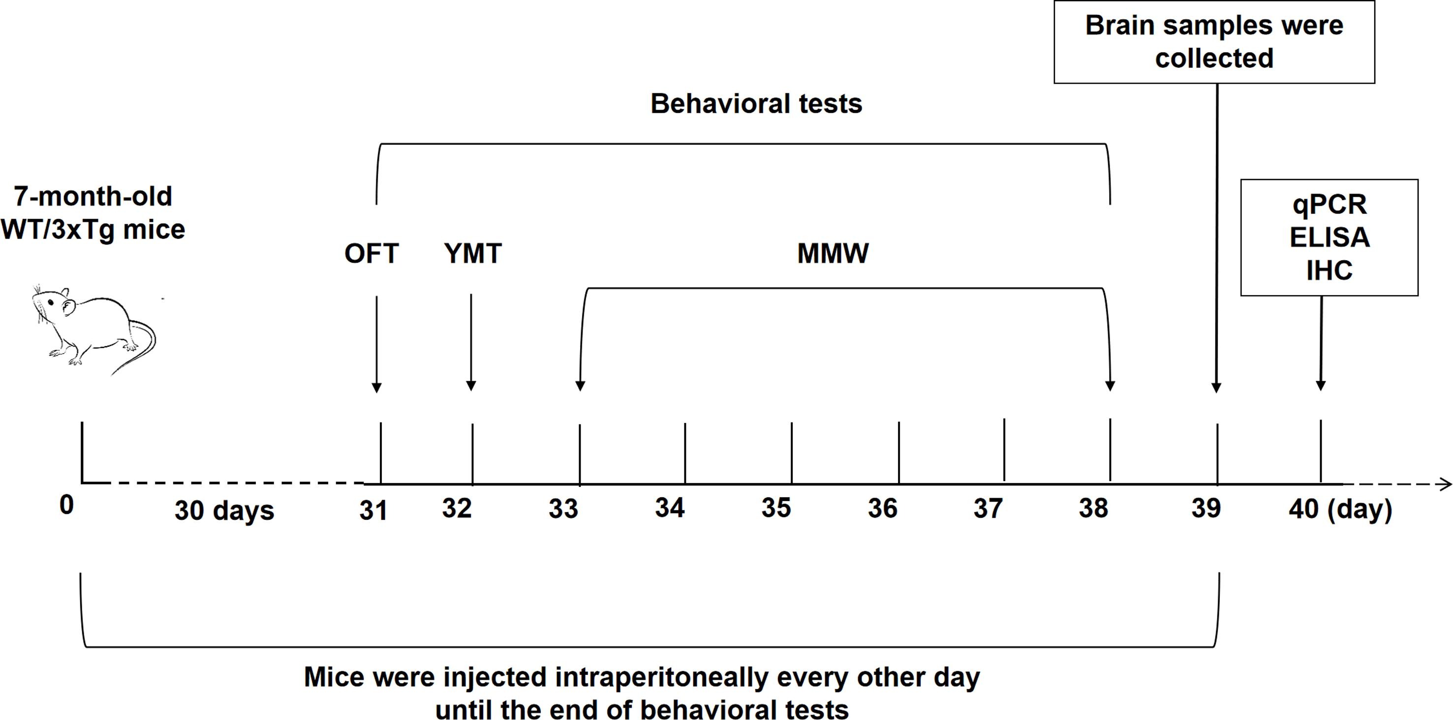
Zhao-Jun Wang and colleagues studied semaglutide in 7-month-old APP/PS1/tau transgenic (3xTg) mice and wild-type mice. Following the study-specified exposure period, the investigators performed behavioral testing and molecular analyses of brain tissue. The authors reported differences in working-memory and spatial-reference-memory measures in 3xTg mice, together with changes in cortical and hippocampal inflammatory mediators and lower Aβ deposition in the hippocampal CA1 region. In accompanying BV2 cell experiments, semaglutide exposure was associated with changes in Aβ1-42-induced apoptosis and markers used to distinguish M1- and M2-like microglial phenotypes.[2]

Fig1. Experimental timeline for the APP/PS1/tau mouse study reported by Wang et al. OFT: open field test; YMT: Y-maze test; MWM: Morris water maze; qPCR: quantitative real-time PCR; ELISA: enzyme-linked immunosorbent assay; IHC: immunohistochemistry.[2]

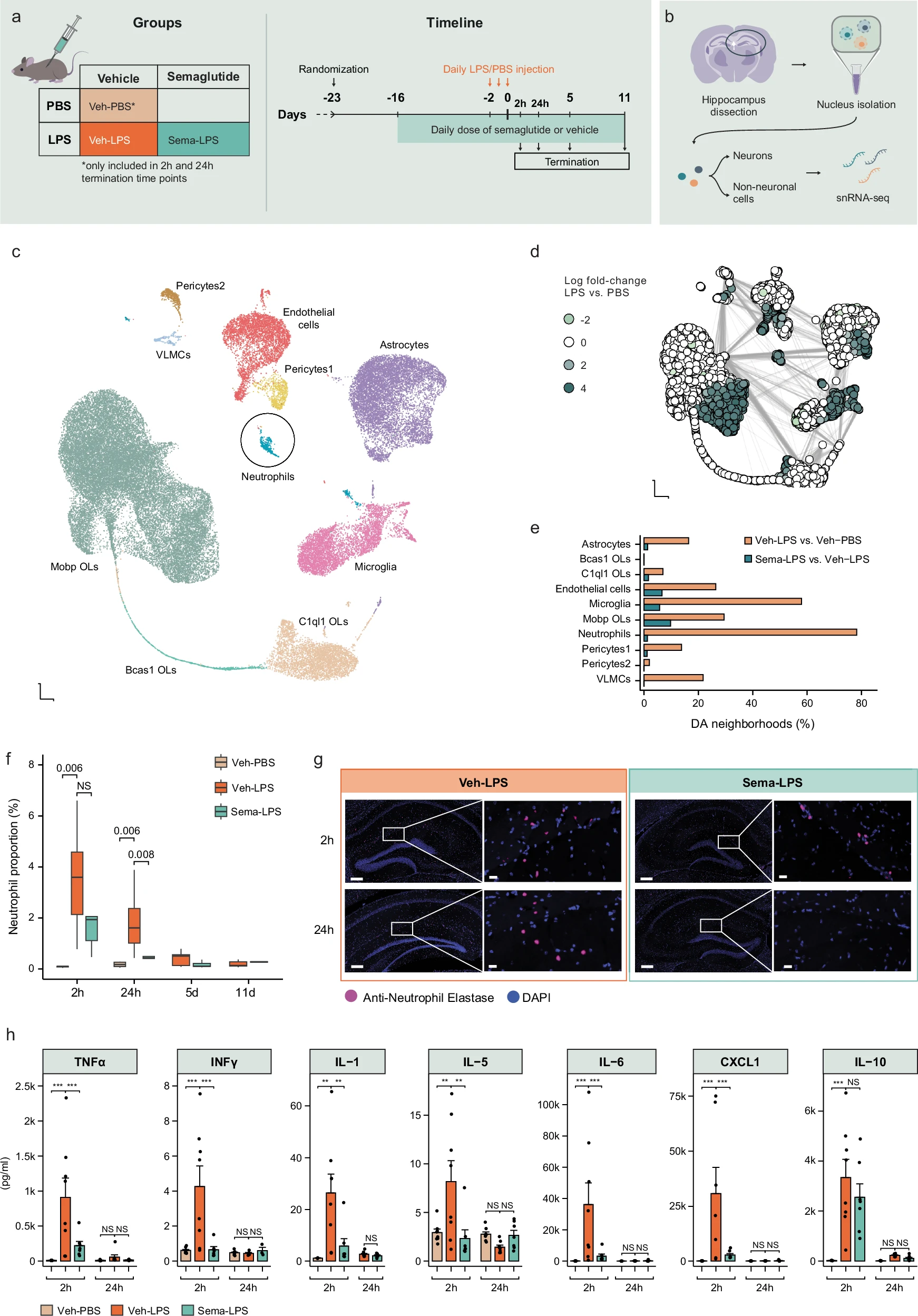
Fig2. Hippocampal cell-type and peripheral inflammatory measurements reported in the LPS mouse model studied by Belmont-Rausch et al.[3]
a, Overview of the in vivo study. b, Overview of the hippocampal snRNA-seq protocol. c-e, Cell-type and differential-abundance analyses of hippocampal non-neuronal cells. f-g, Neutrophil measurements in the hippocampus. h, Terminal plasma measurements of TNFα, IFNγ, IL-1β, IL-5, IL-6, CXCL1, and IL-10. Group sizes, time points, statistical tests, and complete experimental conditions are reported in the original publication.[3]
Vincenzo Quagliariello and colleagues reported a conference-abstract study using a non-diabetic mouse model exposed sequentially to doxorubicin and trastuzumab. Cardiac function was assessed by echocardiography, while histological and circulating markers were used to examine myocardial and inflammatory changes. The authors reported differences in left ventricular ejection fraction, strain measurements, myocardial fibrosis and hypertrophy, troponin, and inflammatory mediators in the semaglutide study group compared with the vehicle group under this experimental protocol.[4]
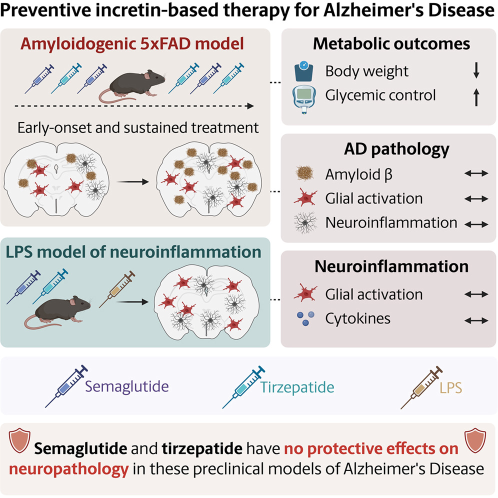
Anika Vear and colleagues examined semaglutide and GLP-2 in 5xFAD mice treated for 2 or 4 months. Both compounds produced metabolic changes in the model, but the authors did not observe measurable differences in memory or learning tasks, amyloid-β plaque deposition, or glial activation. In a separate non-amyloidogenic LPS model, short-term incretin pretreatment did not alter microglial activation or measured inflammatory markers. The authors concluded that these preclinical experiments did not show a slowing of neuropathological progression in the models studied.[5]

Fig3. Graphical overview of the 5xFAD and LPS preclinical experiments reported by Vear et al.[5]
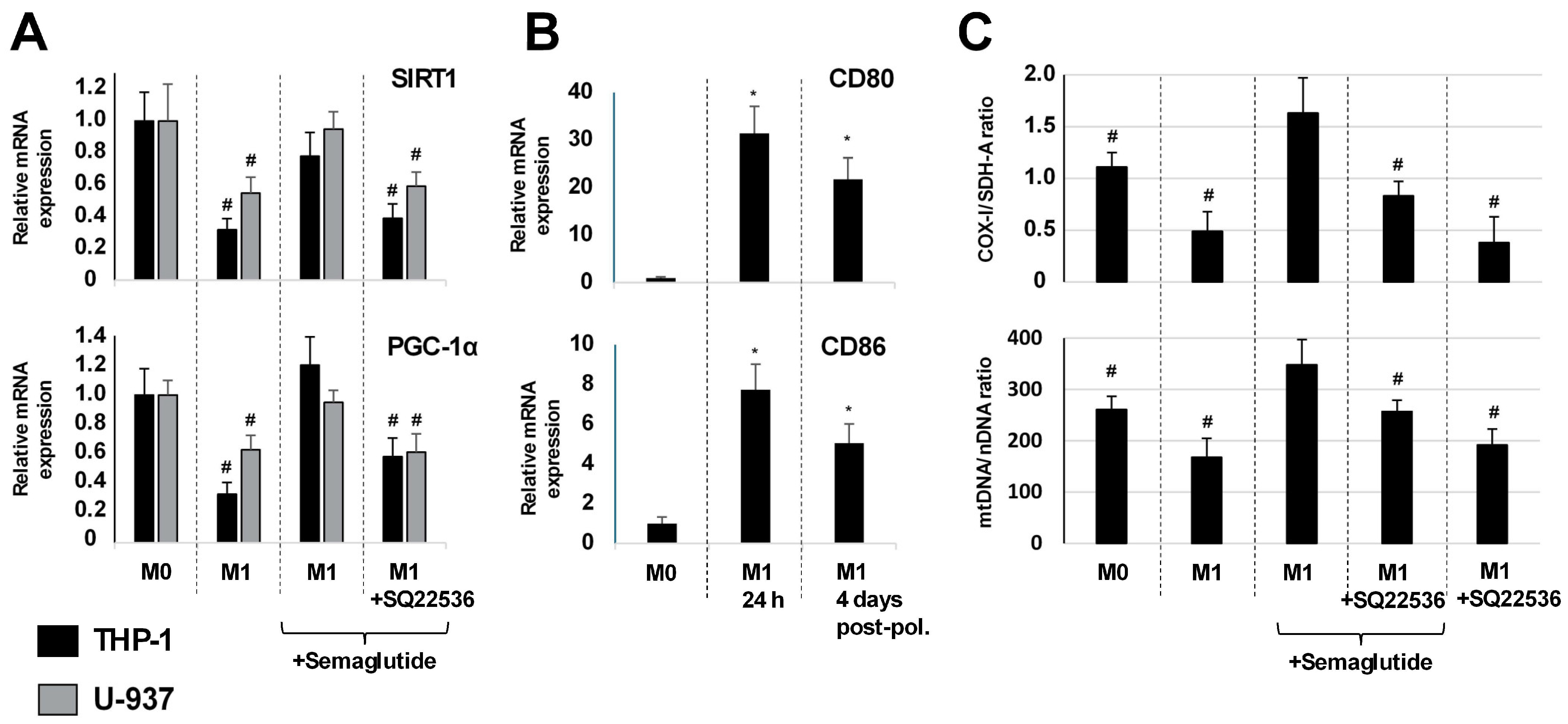
Vsevolod V. Pavshintsev and colleagues differentiated THP-1 and U-937 monocyte cell lines into macrophage-like cells and used LPS/IFN-γ to generate an M1-like polarization model. Cells were exposed to semaglutide with or without the adenylyl cyclase inhibitor SQ22536. The investigators measured intracellular cAMP, macrophage-associated markers, cytokines, SIRT1 and PGC-1α expression, and COX-1/SDH-A and mtDNA/nDNA ratios. The authors reported cAMP accumulation after semaglutide exposure, changes in inflammatory and M2-associated markers, and changes in measurements associated with mitochondrial biogenesis. These responses were attenuated by SQ22536, supporting a contribution of cAMP signaling under the cell-culture conditions used in the study.[6]

Fig4. SIRT1, PGC-1α, macrophage-marker, and mitochondrial measurements reported in THP-1- and U-937-derived macrophage-like cells exposed to semaglutide in the cited study.[6]
(A)The source study measured SIRT1 and PGC-1α mRNA expression, CD80 and CD86 expression, COX-1/SDH-A protein ratios, and mtDNA/nDNA ratios in THP-1- and U-937-derived macrophage-like cells under M0-like and LPS/IFN-γ-polarized conditions. Semaglutide and SQ22536 were used according to the experimental design described by the authors. Sample sizes, concentrations, normalization methods, and statistical comparisons are reported in the original publication.[6]
Research Limitations
Takizawa and colleagues examined semaglutide in Caco-2 intestinal epithelial cells at 5, 50, and 500 ng/mL. Under the conditions tested, semaglutide did not reduce cell proliferation or viability and produced relatively limited changes in the epithelial-barrier markers examined. Because this work used a single in vitro epithelial cell model, it does not establish intestinal effects in vivo and should not be extrapolated beyond the experimental system.[7]
Preclinical findings involving semaglutide are not consistent across neuroinflammatory and neurodegenerative models. The APP/PS1/tau study reported behavioral, inflammatory, and amyloid-associated differences after semaglutide exposure, while the 5xFAD study did not detect changes in memory, amyloid-β plaque deposition, or glial activation under its experimental conditions.[2,5] The LPS study by Belmont-Rausch et al. examined acute neuroinflammatory responses rather than Alzheimer’s disease progression.[3] Differences in mouse strain, disease model, study duration, endpoints, and experimental design limit direct comparison among these studies and do not establish clinical effects in humans.
COCER Peptides presents Semaglutide solely for laboratory research and analytical use. It is not intended for human or veterinary use. The research described above summarizes findings reported in third-party publications and is not a statement of clinical efficacy, safety, diagnosis, treatment, prevention, or an instruction for use.
▎Relevant Citations
References
[1] Papakonstantinou I, Tsioufis K, Katsi V. Spotlight on the Mechanism of Action of Semaglutide[J]. Current Issues in Molecular Biology, 2024,46(12):14514-14541.
DOI:10.3390/cimb46120872.
[2] Wang Z, Han W, Chai S, et al. Semaglutide promotes the transition of microglia from M1 to M2 type to reduce brain inflammation in APP/PS1/tau mice[J]. Neuroscience, 2024,563:222-234.
DOI:10.1016/j.neuroscience.2024.11.022.
[3] Belmont-Rausch D M, Ludwig M Q, Bentsen M A, et al. Semaglutide attenuates neuroinflammation in male mice[J]. Nature Communications, 2026,17:7328.
DOI:10.1038/s41467-026-74038-4.
[4] Quagliariello V, Berretta M, Barbato M, et al. The GLP-1 receptor agonist semaglutide preserves cardiac function, reduces inflammation-driven cardiotoxicity with cardio-renal benefits in non-diabetic models exposed to anthracyclines and HER2 blockade[J]. European Heart Journal Supplements, 2026,28(Supplement_6):suag058.049.
DOI:10.1093/eurheartjsupp/suag058.049
[5] Vear A, Olsen S A, Lange E C H, et al. Preventative semaglutide and GLP-2 treatment does not alter disease progression in the 5xFAD mouse model of Alzheimer's disease[J]. Cell Reports Medicine, 2026,7(7):102906.
DOI:10.1016/j.xcrm.2026.102906.
[6] Pavshintsev V V, Erbaeva A Z, Vatlin A A, et al. Semaglutide Attenuates Inflammatory Macrophage Polarization and Promotes Changes Associated with Mitochondrial Biogenesis Through cAMP-Dependent Signaling[J]. Biomedicines, 2026,14(9):1987.
DOI:10.3390/biomedicines14091987.
[7] Takizawa Y, Kato A, Onsui A, et al. Impact of Glucagon-like Peptide-1 Receptor Agonists on Intestinal Epithelial Cell Barrier[J]. European Pharmaceutical Journal, 2024,71(1):43-52.
DOI:10.2478/afpuc-2024-0008.
[8] NCBI PubChem. Semaglutide. PubChem CID 56843331.
ALL PRODUCTS, ARTICLES, TECHNICAL MATERIALS, PRODUCT DESCRIPTIONS, AND OTHER INFORMATION PROVIDED ON THIS WEBSITE ARE INTENDED SOLELY FOR LEGITIMATE LABORATORY RESEARCH, INFORMATIONAL, AND EDUCATIONAL PURPOSES.
Products offered by Cocer Peptides are intended exclusively for in vitro and laboratory research and are NOT intended for human or veterinary use, human consumption, clinical use, diagnostic use, therapeutic use, compounding, self-administration, injection, or any other form of administration to humans or animals. These products are not intended to diagnose, treat, cure, mitigate, or prevent any disease or medical condition, nor are they intended to affect the structure or function of the human or animal body. Unless expressly stated otherwise, products offered on this website have not been evaluated or approved by the U.S. Food and Drug Administration (FDA) for human or veterinary use. Any references to published studies, biochemical pathways, molecular mechanisms, or experimental findings are provided solely for scientific discussion and research purposes and do not constitute medical advice, therapeutic claims, dosing guidance, or instructions for use. Purchasers are solely responsible for ensuring that all products are acquired, handled, stored, used, and disposed of in accordance with applicable laws, regulations, institutional requirements, and laboratory safety standards.