
|


1kits(10Vials)
| Quantity: | |
|---|---|
▎TB-500 Structure
Source: PubChem |
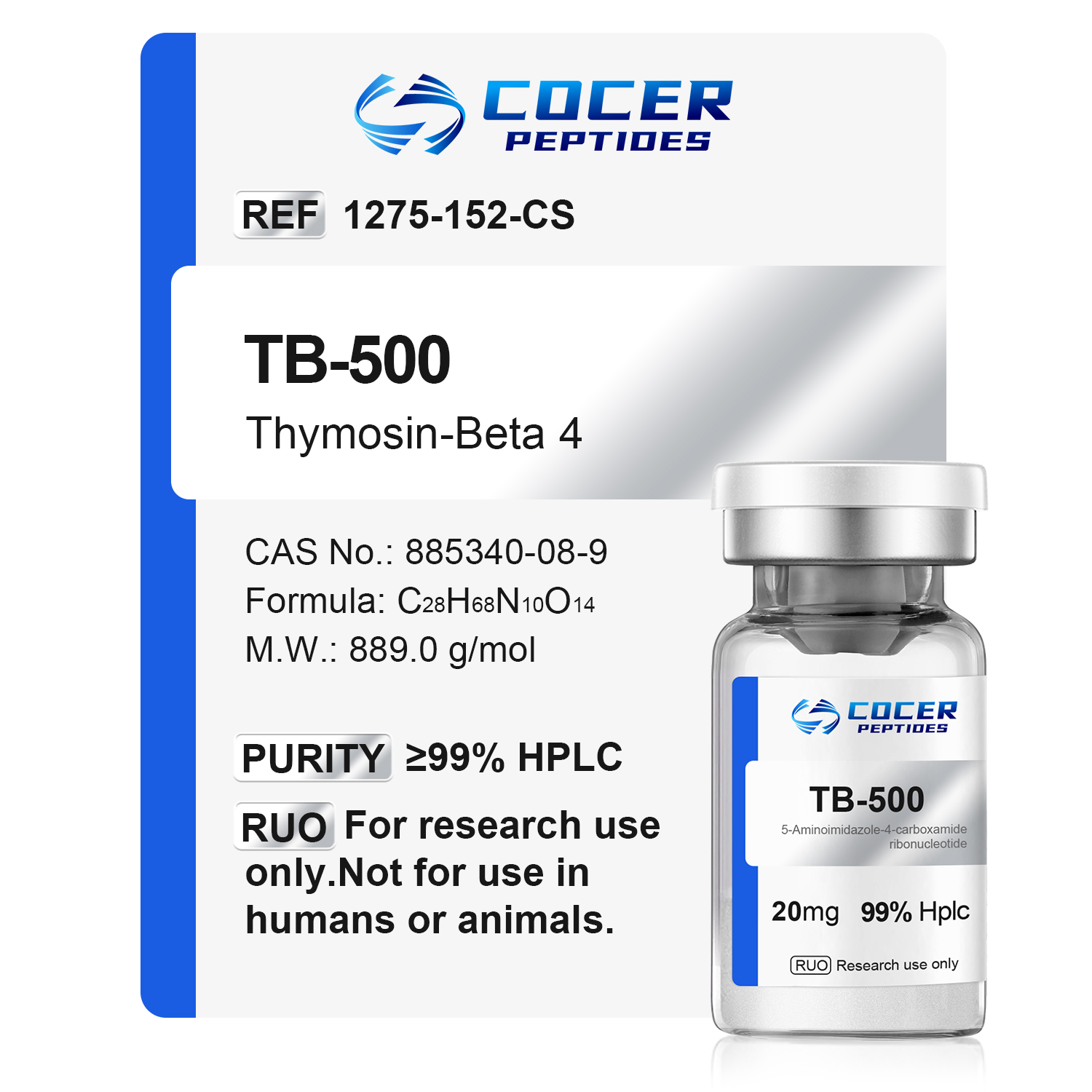
Sequence: LKKTETQ Molecular Formula: C38H68N10O14 Molecular Weight: 889.0 g/mol CAS Number: 885340-08-9 PubChem CID: 62707662 Synonyms: QHK6Z47GTG |
▎TB-500 Research
Molecular Background
TB-500 is an N-terminally acetylated heptapeptide with the sequence Ac-LKKTETQ, corresponding to residues 17–23 of thymosin beta-4 (Tβ4). TB-500 is distinct from full-length Tβ4, which is a 43-amino-acid peptide. PubChem lists TB-500 under CID 62707662, with the molecular formula C38H68N10O14 and a molecular weight of approximately 889.0 g/mol.[1,8]
Research Progress
Researchers led by Khandoker Asiqur Rahaman used ultra-high-performance liquid chromatography coupled with Q-Exactive Orbitrap mass spectrometry to develop a method for the simultaneous analysis of TB-500 and its metabolites. The investigators examined TB-500 metabolism in human serum, in vitro enzyme systems, and urine collected from rats exposed to TB-500. Ac-LK was reported as the major early metabolite, while Ac-LKK remained detectable for a longer period. In fibroblast scratch assays, the parent TB-500 did not show a significant wound-closure response compared with control, whereas the metabolite Ac-LKKTE showed a significant difference under the assay conditions.[1]
The following studies investigated full-length or recombinant thymosin beta-4 (Tβ4), rather than TB-500 itself. They are included as related Tβ4 research and should not be interpreted as direct evidence for the N-acetylated TB-500 heptapeptide.[7]
Wandi Li’s team isolated adipose-derived stem cells (ADSCs) from patients who had undergone liposuction and exposed the cells to full-length Tβ4 in vitro. The investigators assessed proliferation, apoptosis, migration, and gene-expression endpoints using CCK-8 assays, flow cytometry, scratch assays, and RT-qPCR. They reported higher ADSC proliferation at selected Tβ4 concentrations and changes in proliferation- and apoptosis-related measurements under the study conditions.[2] Xiaoyu Zhang and colleagues subsequently studied full-length Tβ4 in an ADSC/fat-graft experimental system and reported increased tunneling nanotube formation through the Rac/F-actin pathway, together with changes in mitochondrial transfer, oxidative-stress markers, apoptosis, and vascularization-related endpoints.[3]
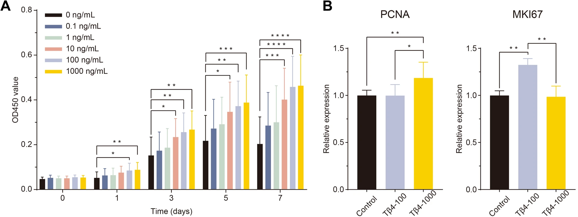
Fig1. Proliferation-related measurements reported in adipose-derived stem cells exposed to full-length Tβ4 in the cited study.[2]
A CCK-8 assay was used to compare ADSC proliferation across Tβ4 concentrations and time points. B RT-qPCR was used to evaluate proliferation-related gene expression, including PCNA and MKI67. Statistical comparisons and complete experimental conditions are reported in the original publication.[2]
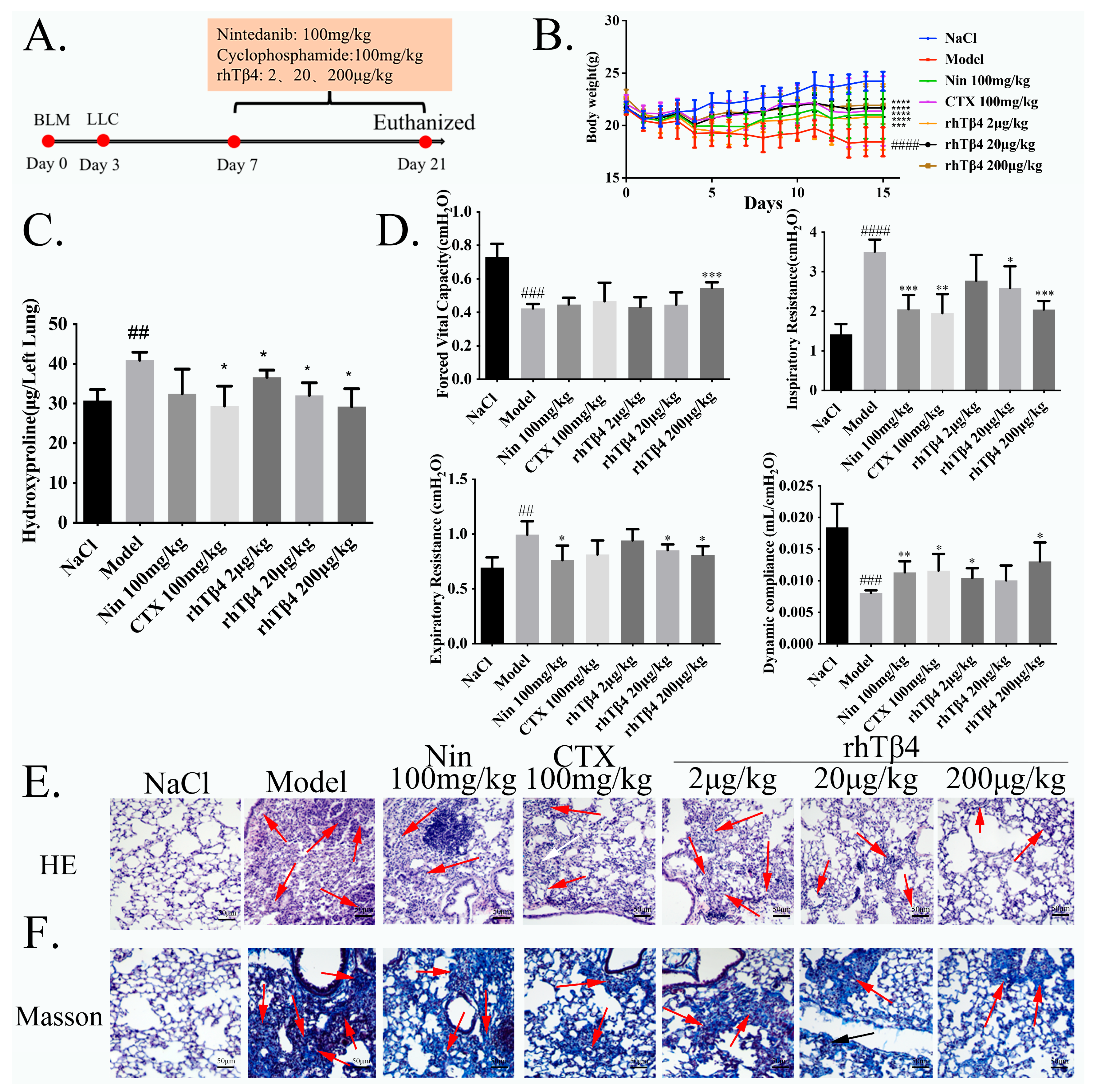
Fig2. Lung-function, histological, and fibrosis-related measurements reported after recombinant human Tβ4 exposure in the bleomycin-based mouse model.[4]
The source figure shows the experimental schedule, body-weight measurements, lung hydroxyproline content, lung-function parameters, and histological staining in the reported mouse groups. Group definitions, study doses, statistical methods, and complete experimental procedures are provided in the original publication.[4]
Yuze Zhang and colleagues studied recombinant human thymosin beta-4 (rhTβ4) in mouse models of cardiac ischemia-reperfusion and permanent coronary ligation. The authors reported differences in cardiac-function, fibrosis, NT-proBNP, and infarct-related measurements in rhTβ4 study groups. RNA-sequencing and cell-based hypoxia/reoxygenation experiments were also used to investigate ErbB-associated signaling, including ErbB2/Raf1 and apoptosis-related endpoints. This work concerned recombinant full-length Tβ4 and should not be treated as direct TB-500 evidence.[5]
Xiankui Li and colleagues evaluated recombinant human thymosin beta-4 using spleen-cell proliferation assays and a full-thickness cutaneous wound model in BALB/c mice. The authors reported changes in lymphocyte proliferation and differentiation and in wound-closure measurements under the conditions of the mouse study. The material studied was recombinant full-length Tβ4, not TB-500.[6]
Research Limitations
Direct published evidence on TB-500 itself remains limited. Rahaman et al. characterized the metabolism of the N-acetylated heptapeptide and reported several shorter metabolites, including Ac-LK and Ac-LKK. In the fibroblast scratch assay used in that study, the parent TB-500 did not show significant wound-closure activity compared with control, while Ac-LKKTE did.[1] These findings do not establish the biological activity of TB-500 in other experimental systems.
An additional limitation is the frequent use of the names TB-500 and thymosin beta-4 as though they describe the same molecular entity. They do not. A 2026 scoping review found that most of the published repair-related literature concerns full-length Tβ4, while direct TB-500 evidence was limited to a single included study.[7] Results obtained with synthetic or recombinant full-length Tβ4 therefore should not be automatically extrapolated to the seven-amino-acid TB-500 fragment.
COCER Peptides presents TB-500 solely for laboratory research and analytical use. It is not intended for human or veterinary use. The research described above summarizes findings reported in third-party publications and distinguishes direct TB-500 studies from related full-length Tβ4 studies; it is not a statement of clinical efficacy, safety, diagnosis, treatment, prevention, or an instruction for use.


